The next follow-up visit should not exceed 4 months after delivery of the crown. In the four-hour MA clinic session participants will take a written exam and take satisfactory periapical and bitewings using digital sensors on a manikin.

Periapical Radiography Pocket Dentistry
Furthermore we will be free to use any ideas concepts know-how or techniques contained in any communication you send to us for any purpose whatsoever including but not limited to developing manufacturing and marketing products using such information.

. A record peri-apical X-ray after delivery of the final prosthesis is necessary at this point. This radiograph will be useful for follow-up and maintenance comparisons of bone level with later radiographs Figures 25a- -c. At the same time MRI as a non-invasive diagnostic tool in apical periodontitis 68 is more accurate in this regard and gives a better estimation on the proximity of the lesion to nearby structures 70.
They will become familiar with processing and mounting of films and self-analyze radiographic technical and processing errors. X-ray-based methods have shortcomings and limited performance in measuring the accurate lesion border.

Periapical Radiography Pocket Dentistry
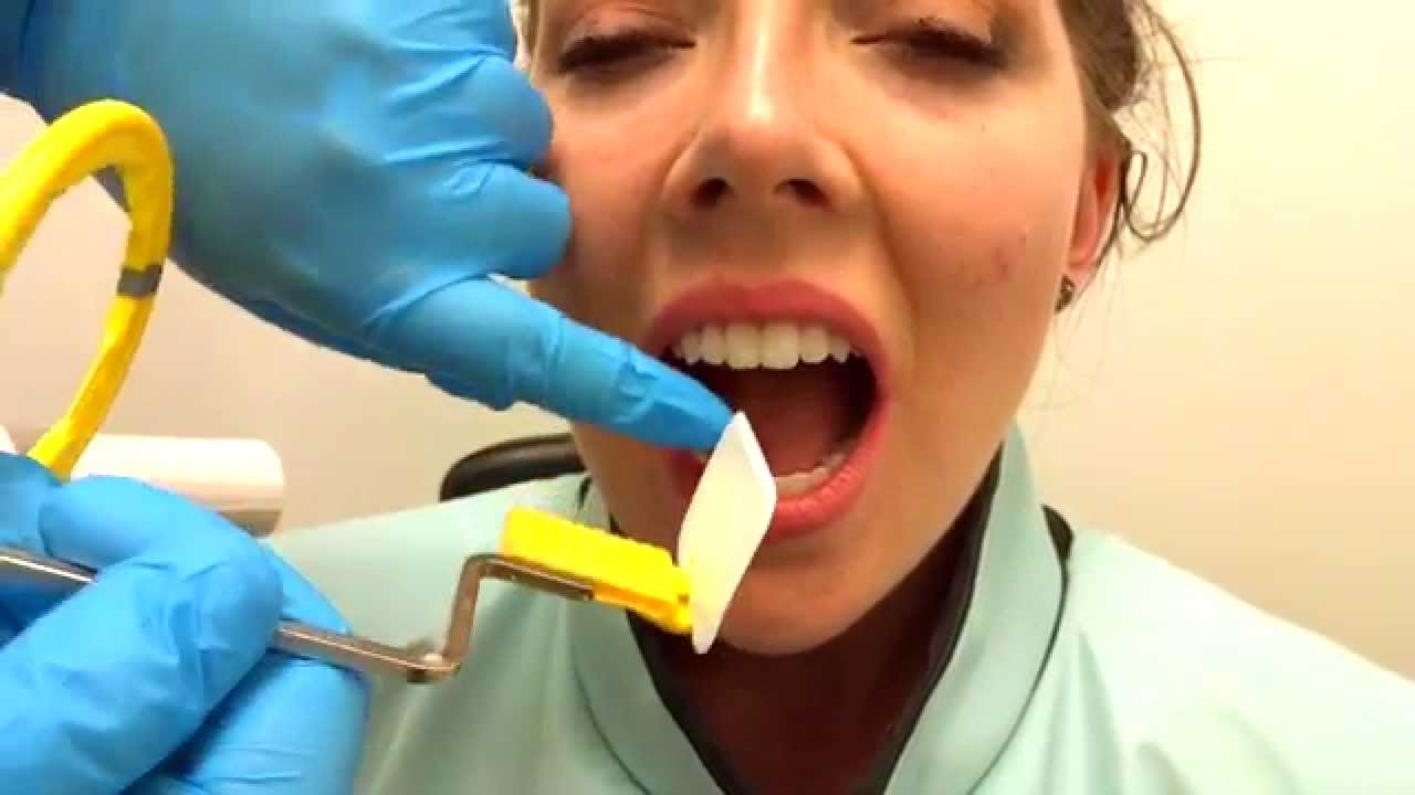
How To Take Periapical Radiographs Youtube

Periapical Radiography Pocket Dentistry
Periapical Radiography Pocket Dentistry

How Make Periapical X Ray

Periapical Radiography Pocket Dentistry

Periapical Radiography Pocket Dentistry

Periapical Radiography Pocket Dentistry
0 comments
Post a Comment